Hot NewsPosterior Rib Cage Muscles - Anatomy Of The Thoracic Wall Pulmonary Cavities And Mediastinum Thoracic Key
Posterior Rib Cage Muscles - Anatomy Of The Thoracic Wall Pulmonary Cavities And Mediastinum Thoracic Key
Posterior Rib Cage Muscles - Anatomy Of The Thoracic Wall Pulmonary Cavities And Mediastinum Thoracic Key. There are some other muscles that do not comprise the thoracic wall, but do attach to it. Both the rib cage and the pelvis are important units of body structure; Muscle anatomy diagram 12 photos of the muscle anatomy diagram canine muscle anatomy diagram, dog muscle anatomy diagram, lower leg muscle anatomy diagram, muscle anatomy of human back, tricep muscle anatomy diagram, human muscles, canine muscle anatomy diagram, dog muscle anatomy diagram, lower leg muscle anatomy diagram. On the interior wall of the rib body is a channel, sulcus costae, with blood vessels and nerves. Costochondritis or tietze's syndrome is another common cause of rib cage pain.
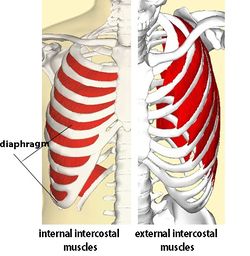
Posterior rib cage muscles : There are some other muscles that do not comprise the thoracic wall, but do attach to it. Rib 2 is thinner and longer than rib 1, and has two articular facets on the head as normal. Of all 24 ribs, the first seven pairs are often labeled as 'true.'. The intercostals (external, internal and innermost), subcostals, and transversus thoracis.
The Respiratory Muscles Structure And Function Organization Of The Respiratory System from www.pharmacy180.com Other times, the cause of rib pain is more serious, such as appendicitis, pancreatitis, lung cancer, a pulmonary embolism, gallbladder problems, fibromyalgia, or heart attacks and other heart problems. Related posts of rib cage diagram with organs woman stomach anatomy. The transversus thoracic muscles originate from the posterior surface of the xiphoid process and the lower part of the body of the sternum. The intermediate muscles of the posterior contribute to movements of the ribcage during respiration. In those cases, deep tissue therapy to release tension in the back may help release the posterior ribs or compensation patterns. Irritation, inflammation, back rib injury, strained or pulled back muscles, or a herniated disc can all cause pain in ribs and back. The configuration of the lower five ribs gives freedom for the expansion of the lower part of the rib cage and for the movements of the diaphragm, which has an extensive origin from the rib cage and the vertebral column. These muscles act to change the volume of the thoracic cavity during respiration.
When the ribcage is fixed contraction results in a posterior pelvic tilt.
This muscle assists in depression of the ribs. Oftentimes, pain under the rib cage is not serious and may be associated with minor conditions like indigestion, gas troubles, or strained muscles. In those cases, deep tissue therapy to release tension in the back may help release the posterior ribs or compensation patterns. Rib cage pain can be associated with bruising, difficulty taking a deep breath, joint pain, and more. Rib cage pain may be sharp, dull, or achy and felt at or below the chest or above the navel on either side. Remember, each rib is like a half moon shape having an anterior, lateral and posterior side. Costochondritis or tietze's syndrome is another common cause of rib cage pain. Find out what functions your muscles perform and. The human rib cage is made up of 12 paired rib bones; The intercostal muscles of the ribcage. The ribs form the main structure of the thoracic cage protecting the thoracic organs, however their main function is to aid respiration.; The intercostals (external, internal and innermost), subcostals, and transversus thoracis. Ribcage aka thoracic cage bony & cartilaginous structure which surrounds thoracic cavity.
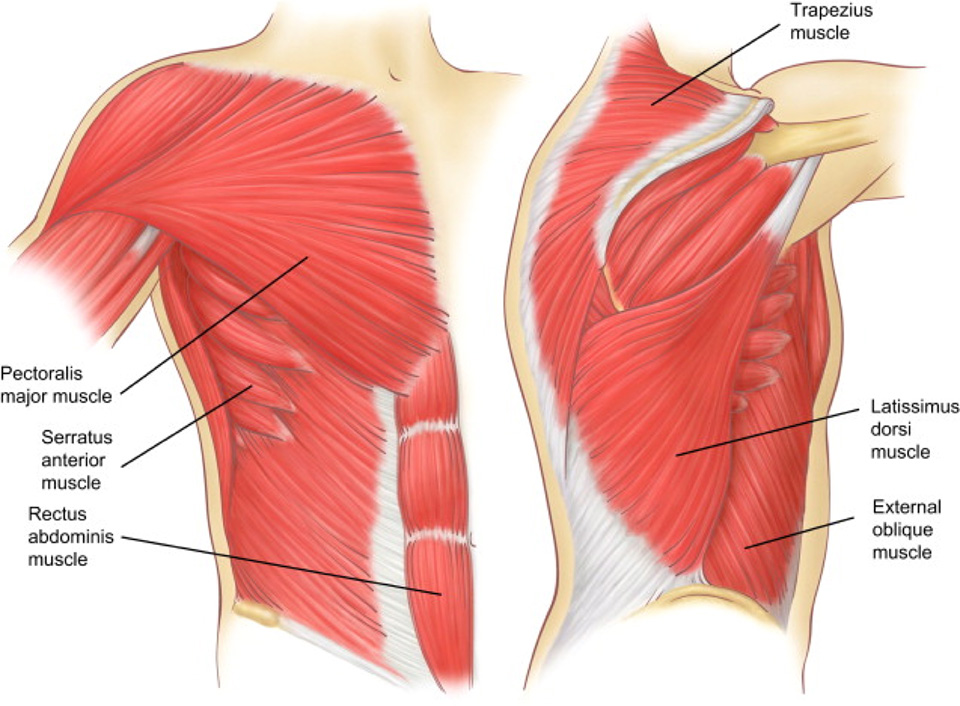
The major abdominal muscles include the transverse abdominals, the rectus abdominis, and the external and internal oblique muscles. If all these muscles are tight, it can leave you feeling constricted. The ribs form the main structure of the thoracic cage protecting the thoracic organs, however their main function is to aid respiration.; Our ribcage exists to protect the heart and lungs. Woman stomach anatomy 7 photos of the woman stomach anatomy activate javascript anatomy of a woman body, female abdomen anatomy, female organ anatomy, human stomach anatomy, stomach anatomy and physiology, stomach anatomy antrum, stomach anatomy pictures, womens stomach anatomy, stomach, anatomy of a woman body, female.
Intercostal Muscle Strain Physiopedia from www.physio-pedia.com There are some other muscles that do not comprise the thoracic wall, but do attach to it. The configuration of the lower five ribs gives freedom for the expansion of the lower part of the rib cage and for the movements of the diaphragm, which has an extensive origin from the rib cage and the vertebral column. These muscles act to change the volume of the thoracic cavity during respiration. Remember, each rib is like a half moon shape having an anterior, lateral and posterior side. Read more below to learn what may be causing your rib pain and when to seek treatment. They wrap around your chest between your ribs and are attached to the sternum and. When the ribcage is fixed contraction results in a posterior pelvic tilt. Rib cage pain can be associated with bruising, difficulty taking a deep breath, joint pain, and more.
These muscles act to change the volume of the thoracic cavity during respiration.
The ribs form the main structure of the thoracic cage protecting the thoracic organs, however their main function is to aid respiration.; If all these muscles are tight, it can leave you feeling constricted. The other attachment of these muscles is usually considered to be either superior or inferior to the rib attachment. An exception to this rule is that the first rib articulates with the first. When the ribcage is fixed contraction results in a posterior pelvic tilt. The rib cage intrinsically holds the muscles of respiration (diaphragm, intercostal muscles, etc.) that are crucial for active inhalation and forced exhalation, and therefore has a major ventilatory function in the respiratory system. Both the rib cage and the pelvis are important units of body structure; The configuration of the lower five ribs gives freedom for the expansion of the lower part of the rib cage and for the movements of the diaphragm, which has an extensive origin from the rib cage and the vertebral column. It may occur after an obvious injury or without explanation. These muscles act to change the volume of the thoracic cavity during respiration. It usually occurs in the cartilage. They wrap around your chest between your ribs and are attached to the sternum and. Sudden, severe upper back/rib pain.
Rib cage pain may be sharp, dull, or achy and felt at or below the chest or above the navel on either side. If all these muscles are tight, it can leave you feeling constricted. These muscles may be located anteriorly, posteriorly, and/or laterally. Intercostal muscle strain is an injury affecting the muscles between two or more ribs. Posterior rib cage muscles :
Costochondritis Chest Wall Pain Rib Injury Clinic from www.ribinjuryclinic.com The rib cage intrinsically holds the muscles of respiration (diaphragm, intercostal muscles, etc.) that are crucial for active inhalation and forced exhalation, and therefore has a major ventilatory function in the respiratory system. An exception to this rule is that the first rib articulates with the first. The middle and upper part of your spine is called the thoracic region and it helps to support your upper body. Each rib articulates posteriorly with two thoracic vertebrae by the costovertebral joint. The ribs are the bony framework of the thoracic cavity. Muscle spasms felt within the rib cage may also be caused by the abdominal muscles. The human rib cage is made up of 12 paired rib bones; Posterior rib cage muscles :
These muscles act to change the volume of the thoracic cavity during respiration.
Other times, the cause of rib pain is more serious, such as appendicitis, pancreatitis, lung cancer, a pulmonary embolism, gallbladder problems, fibromyalgia, or heart attacks and other heart problems. Posterior rib cage muscles : The middle and upper part of your spine is called the thoracic region and it helps to support your upper body. The human rib cage is made up of 12 paired rib bones; Intercostal muscle strain is an injury affecting the muscles between two or more ribs. Oftentimes, pain under the rib cage is not serious and may be associated with minor conditions like indigestion, gas troubles, or strained muscles. There are five muscles that make up the thoracic cage; The intercostal muscles have different layers that are attached to the ribs to help build the chest wall and. Each are symmetrically paired on a right and left side. Remember, each rib is like a half moon shape having an anterior, lateral and posterior side. Of all 24 ribs, the first seven pairs are often labeled as 'true.'. The intermediate muscles of the posterior contribute to movements of the ribcage during respiration. These muscles act to change the volume of the thoracic cavity during respiration.
The configuration of the lower five ribs gives freedom for the expansion of the lower part of the rib cage and for the movements of the diaphragm, which has an extensive origin from the rib cage and the vertebral column rib cage muscles. An exception to this rule is that the first rib articulates with the first.

0 comments